Heart Test Echocardiogram: What to Expect and Why It Matters
An echocardiogram is a key test to understand your heart’s health. It uses sound waves to create detailed images of your heart, showing how blood flows and how well your heart valves work. This non-invasive procedure offers a clear picture of your heart’s condition, helping doctors diagnose various issues like heart disease or valve problems.

You might wonder why this test is essential. Echocardiograms provide vital information that can reveal conditions you might not even feel, like subtle heart valve issues or early heart failure signs. Knowing what’s happening inside your heart can lead to better treatment plans and more peace of mind.
The process of getting an echocardiogram is simple and safe. You lie down while a technician places sensors on your chest to capture the images. The test is usually quick, often taking less than an hour, and you can go about your day right after.
Key Takeaways
- Echocardiograms use sound waves to create images of your heart.
- The test helps diagnose heart conditions early.
- The procedure is quick, safe, and non-invasive.
Understanding Echocardiograms

Echocardiograms use ultrasound to create detailed images of the heart. These images help in diagnosing various heart conditions by showing the heart’s structure, function, and blood flow.

What Is an Echocardiogram?
An echocardiogram, often called an “echo,” is a test that uses sound waves to produce images of your heart. These images help your doctor see your heart’s size, shape, and how well it is functioning.
Ultrasound technology is at the core of this test. Sound waves bounce off your heart and create moving images on a screen. These images show the heart’s muscles, chambers, valves, and blood vessels.
An echocardiogram can also help find problems like blood clots, tumors, and congenital heart defects. It’s a non-invasive, painless test that usually takes about 30 to 60 minutes.
Types of Echocardiograms
There are several types of echocardiograms:
- Transthoracic echocardiogram (TTE): This is the most common type. A device called a transducer is moved across your chest to create images of your heart.
- Transoesophageal echocardiogram (TEE): This involves swallowing a flexible tube with a transducer at the end. It provides more detailed images, especially of the heart’s back side.
- Stress echocardiogram: This test is done before and after exercise to see how your heart works under stress. Sometimes, medication is used if you can’t exercise.
- Fetal echocardiogram: This type is used to check a baby’s heart before birth. It can identify congenital heart defects.
Uses of Echocardiography
Echocardiography is used to diagnose and monitor numerous heart conditions. Your doctor might order an echo to:
- Check for heart disease like coronary artery disease.
- Look at the heart valves to see if they are opening and closing correctly.
- Monitor the heart chambers for issues like enlargement or abnormal movement.
- Measure the heart’s pumping strength, which is crucial in diagnosing heart failure.
- Detect blood clots or tumours in the heart.
- Assess congenital heart defects such as holes in the heart.
In essence, echocardiograms provide a thorough insight into how your heart works, helping your healthcare team make informed decisions about your treatment and care.
Preparing for and Undergoing an Echocardiogram
An echocardiogram is a common heart test that uses sound waves to create images of your heart. This procedure is safe and often provides essential information about your heart’s health.
Before the Procedure
Before your echocardiogram, you’ll need to follow some simple steps to prepare. Your doctor will inform you if you need to fast or avoid certain medications. It’s usually best to wear comfortable clothing, though you might be asked to wear a hospital gown. If you are taking any medicines, ensure you continue unless your doctor advises otherwise.
Sometimes, specific types of echocardiograms require more preparation. For example, if undergoing a transesophageal echocardiogram, you may need to avoid eating and drinking for several hours before the test. Your doctor might also give you a sedative to help you relax during this type of echo.

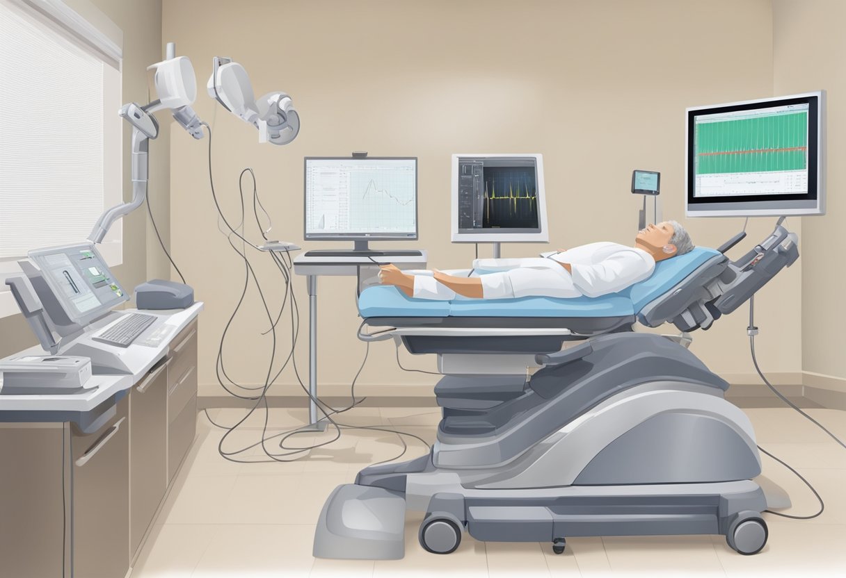
During the Examination
When you arrive at the hospital or clinic, a trained technician or sonographer will guide you through the process. You’ll lie down on an examination table, and the technician will apply a small amount of gel to your chest. This helps the transducer (a hand-held device) to get clear images of your heart.
The technician or sonographer will gently press the transducer against your skin and move it around to capture different views of your heart. If you undergo a stress echocardiogram, you may need to walk on a treadmill or pedal a stationary bike to elevate your heart rate. In a transesophageal echocardiogram, a thin tube with a transducer is guided down your esophagus to get detailed images.
Throughout the test, you might feel a bit of pressure but no significant pain. Electrodes connected to an ECG/EKG machine may be placed on your chest to monitor your heart’s electrical activity during the test.
After the Echo Test
Once the echocardiogram is complete, you can typically return to your normal activities immediately. The gel used on your chest will be wiped off, and any electrodes will be removed. If a sedative was used, you might need someone to drive you home, as the sedative could make you drowsy.
Your doctor will discuss the results with you, usually during a follow-up appointment. You might experience a mild discomfort at the site where the transducer was pressed, but this should go away quickly. If any tube was used during a transesophageal echocardiogram, your throat might feel numb or slightly sore for a short while.
Knowing what to expect can make the experience smoother and less stressful, ensuring you get the most accurate and helpful results from your echocardiogram.
Frequently Asked Questions

Learn more about how echocardiograms are done, why they’re necessary, and what they can reveal about your heart health.
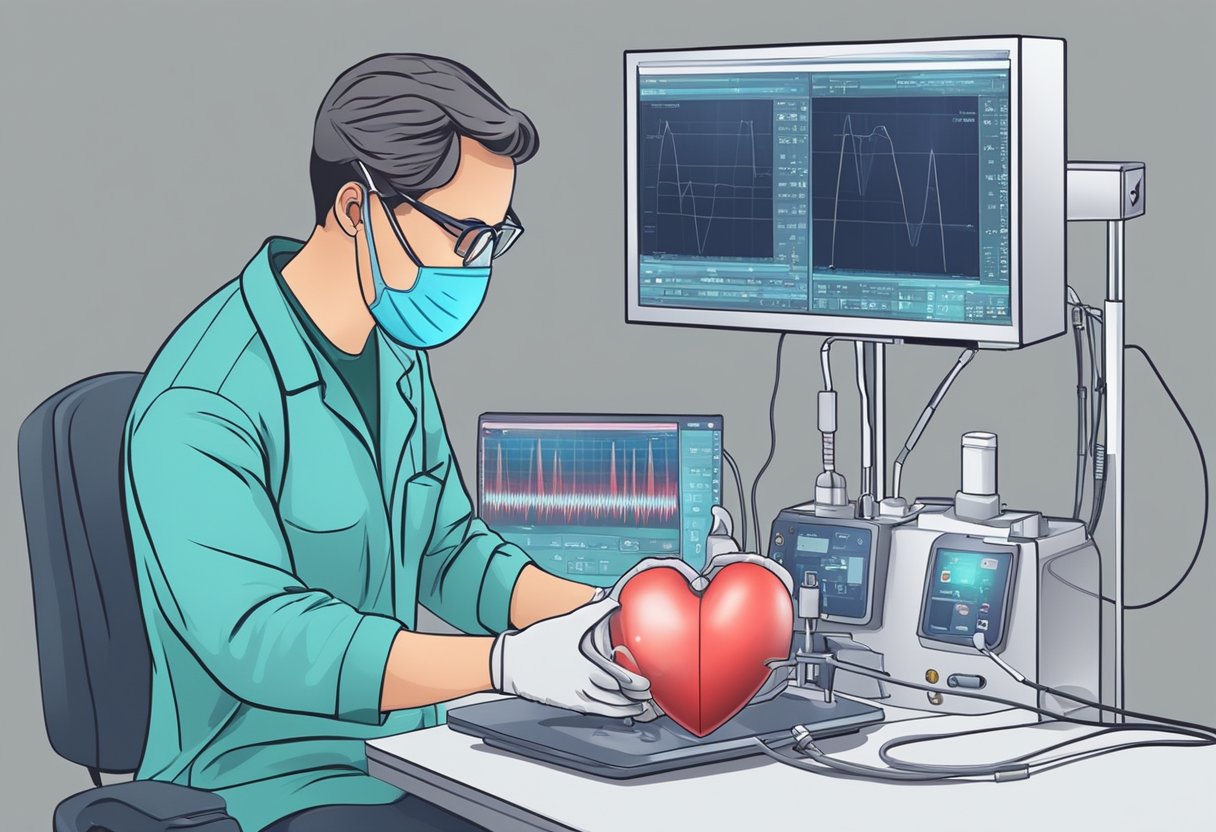
How is an echocardiogram performed?
An echocardiogram is performed using a device called a transducer. This device emits sound waves that bounce off your heart and create images. The transducer is placed on your chest and moved around to capture different views of your heart.
Can a normal echocardiogram ensure that my heart is healthy?
A normal echocardiogram can show that your heart is functioning well at the time of the test. However, it may not detect all heart conditions. It’s important to continue regular check-ups and inform your doctor of any new symptoms.
What are the reasons for conducting an echocardiogram?
Doctors may recommend an echocardiogram if you have symptoms like shortness of breath, chest pain, or irregular heartbeats. It helps in diagnosing conditions such as heart valve problems, heart failure, and other cardiac issues.
What types of abnormalities can an echocardiogram reveal?
An echocardiogram can reveal abnormalities such as problems with the heart valves, heart muscle, and the chambers of the heart. It can also detect issues with blood flow and the presence of congenital heart defects.
What are the necessary preparations before undergoing an echocardiogram?
Generally, no special preparations are needed for a standard echocardiogram. You might be advised to avoid eating or drinking for a few hours if a transesophageal echocardiogram is being done. Always follow your doctor’s instructions.
How much time is typically required for an echocardiogram procedure?
An echocardiogram typically takes about 30 to 60 minutes. The procedure is painless and usually performed on an outpatient basis. After the test, you can usually return to your regular activities immediately.