Heart in Human Body: Essential Engine of Life
The human heart is a fascinating organ, crucial for your survival. It functions as a four-chambered pump that circulates blood throughout your body. This process delivers oxygen and nutrients to your tissues while removing waste products.

Your heart, roughly the size of your fist, is located between your lungs in the middle compartment of your chest. It’s essential to understand how it works, from the rhythmic beats to the flow of blood through its various chambers. By grasping the basics of your heart’s anatomy and function, you can better appreciate its importance and learn how to keep it healthy.
Exploring the structure and function of the heart can also help you recognise symptoms of potential problems. Recognising early signs of heart disease, such as chest pain or shortness of breath, might prompt you to see a doctor sooner, leading to better outcomes.
Key Takeaways
- The heart pumps blood and nutrients throughout your body.
- It is located between your lungs in your chest.
- Recognising heart disease symptoms can improve your health outcomes.
Understanding the Heart
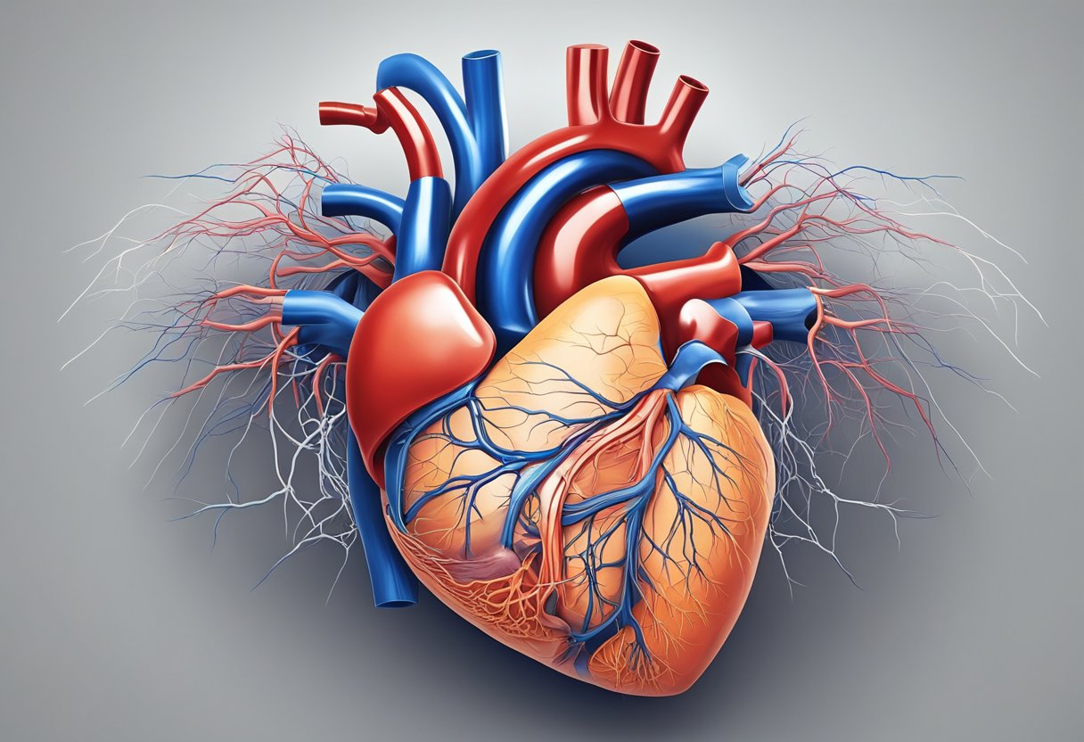
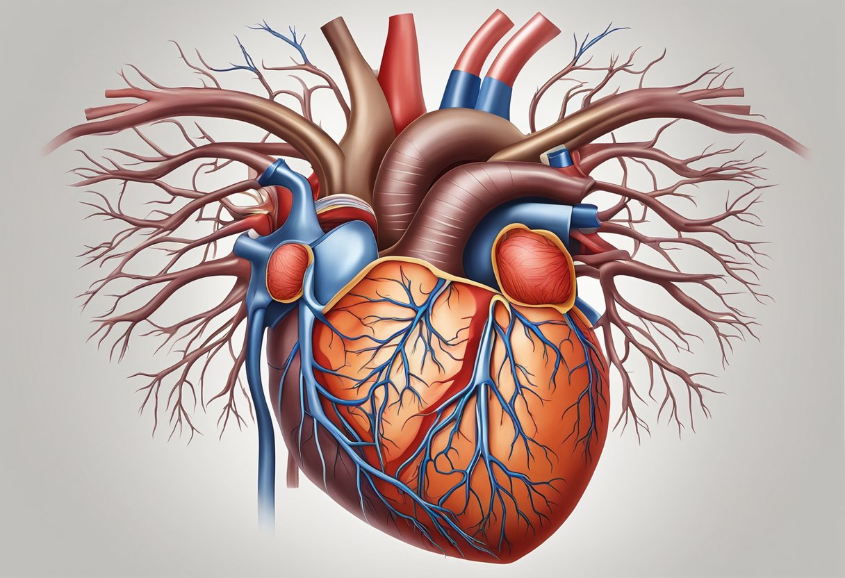
Your heart is a muscular organ that’s essential for pumping blood throughout your body. It has distinct anatomical features and a complex system to ensure it beats correctly.

Cardiac Anatomy and Chambers
Your heart has four primary chambers: two atria and two ventricles. The right atrium receives blood from the body through the venae cavae and pushes it into the right ventricle. The right ventricle then pumps blood to the lungs via the pulmonary trunk.
The left atrium receives oxygenated blood from the lungs and passes it to the left ventricle. The left ventricle, being the strongest chamber, pumps blood to the rest of your body through the aortic valve. The heart’s chambers are separated by atrioventricular (tricuspid and mitral) valves and semilunar valves (aortic and pulmonary) to ensure one-way blood flow.
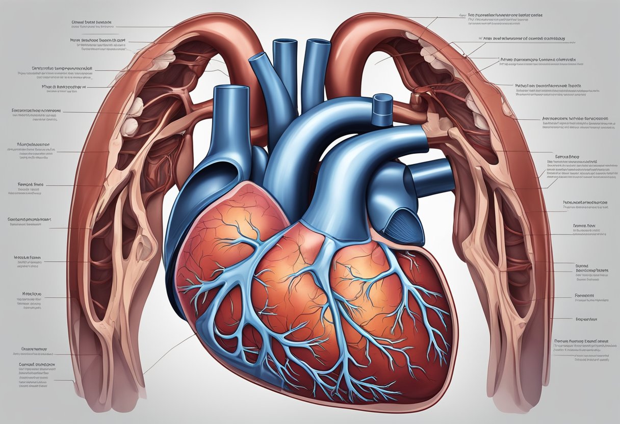
Heart Musculature and Layers
The heart wall consists of three layers: the endocardium, myocardium, and epicardium. The endocardium lines the inside of the heart, while the myocardium is the thick muscle layer responsible for the pumping action. The epicardium covers the outer surface.
Surrounding the heart is the pericardium, a double-layered sac that provides protection and stability. The myocardium contains specialized muscle cells known as cardiac muscle, which contract rhythmically and tirelessly. These muscles work with structures like papillary muscles and pectinate muscles to manage the mechanical stresses during each heartbeat.
The Heart’s Conduction System
Your heart’s rhythm is controlled by its conduction system. The sinoatrial (SA) node, often called the natural pacemaker, initiates each heartbeat. This signal spreads through the atria, causing them to contract and push blood into the ventricles.
The electrical impulse then reaches the atrioventricular (AV) node. After a brief delay, it travels along the bundle of His and into the interventricular septum. The signal continues through the Purkinje fibres, causing the ventricles to contract and pump blood out of your heart. This coordinated sequence ensures a regular and effective cardiac cycle.
The coronary arteries supply oxygen-rich blood to the heart muscle itself. Proper function of this system is crucial to maintaining a healthy heart rate and preventing heart disease.
Circulatory Interactions
The heart is crucial for pumping blood throughout your body, ensuring oxygen and nutrients are delivered to every cell. It maintains a complex network of interactions within the circulatory system to achieve this task.
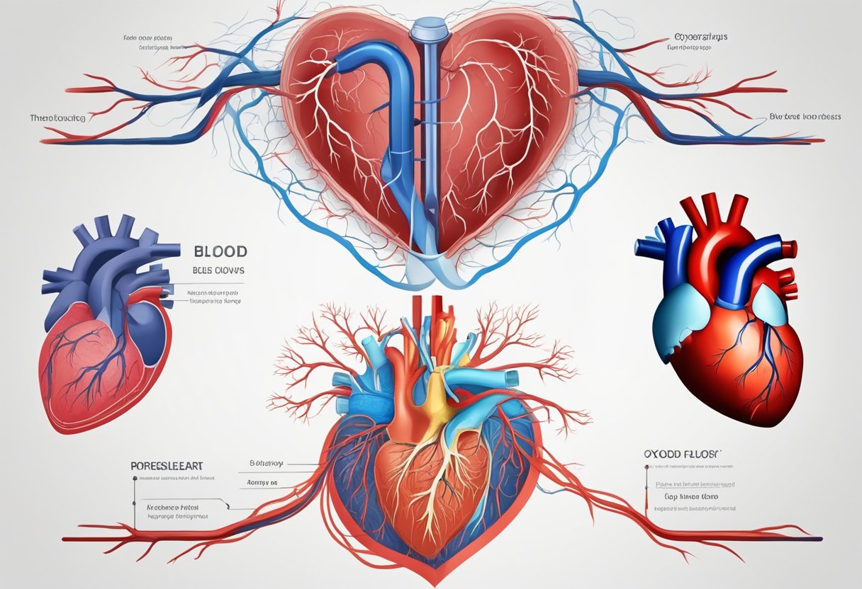
Blood Flow and Vascular System
Blood flows through a network of blood vessels, including arteries, veins, and capillaries. The superior vena cava and inferior vena cava bring deoxygenated blood from your body back to the right atrium of the heart.

From the right atrium, blood moves into the right ventricle, which pumps it to the lungs via the pulmonary artery. In the lungs, the blood picks up oxygen and releases carbon dioxide. Oxygenated blood then returns to the left atrium through the pulmonary veins.
Blood flows into the left ventricle, which pumps it into the aorta. From the aorta, blood travels through arteries to reach various tissues. Capillaries enable nutrient and gas exchange at the cellular level. Veins then carry deoxygenated blood back to the heart. Heart valves, like the bicuspid and tricuspid valves, ensure one-way blood flow, preventing backflow.
Respiratory Connections and Oxygen Exchange
The heart works closely with the lungs to oxygenate blood. Deoxygenated blood enters the lungs via the pulmonary arteries. In the lungs, alveoli are tiny air sacs where gas exchange takes place.
Oxygen passes from the alveoli to the blood, while carbon dioxide is expelled from the blood into the alveoli, ready to be exhaled. The oxygenated blood returns to the left atrium via the pulmonary veins.
This oxygen-rich blood is then pumped from the left ventricle through the aorta to nourish your body. Exercise boosts this process by increasing heart rate and ensuring more oxygen reaches tissues. Monitoring these interactions, healthcare professionals may use a stethoscope to assess heart and lung function.
Frequently Asked Questions
You’re about to learn key facts about the heart’s location, function, and structure, which can help you understand this vital organ better.
Where is the heart positioned in a woman’s body?
In women, the heart is located in the middle of the chest. It sits behind the breastbone and slightly to the left.
How does the heart function?
The heart pumps blood throughout your body. It delivers oxygen and nutrients to your cells and tissues, and removes waste products like carbon dioxide.
Could you show me a diagram of the heart?
A labelled diagram of the heart can help you understand its structure better. [Insert a diagram of the heart here]
What are the key roles of the heart?
The heart’s main roles include pumping blood, delivering oxygen and nutrients, and removing waste products. It also helps maintain blood pressure and circulate hormones.
Does the heart reside on the left side or the right side?
The heart is primarily located in the centre of the chest, veering slightly to the left.
What are the primary chambers of the heart?
The heart has four main chambers: the left atrium, right atrium, left ventricle, and right ventricle. These chambers work together to pump blood efficiently throughout your body.