Cyanotic Congenital Heart Disease Classification: A Simple Guide for Understanding
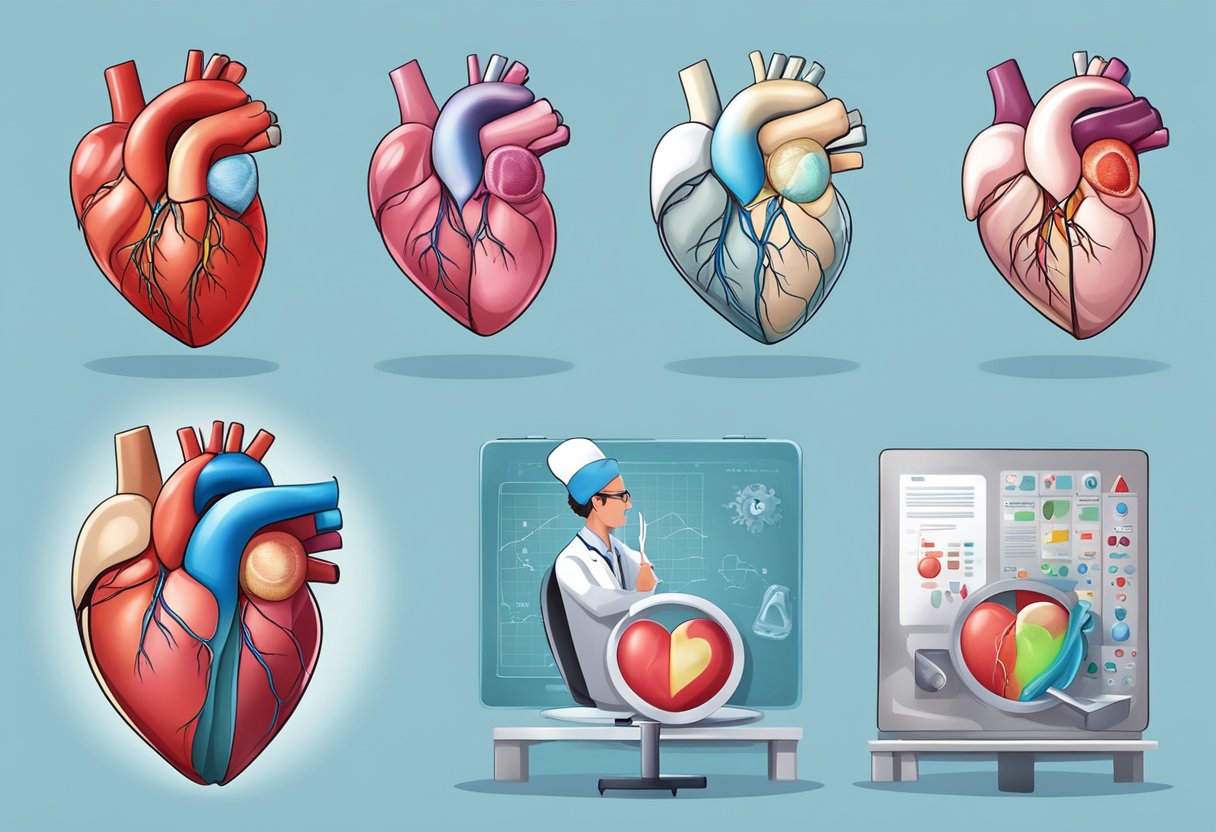
Cyanotic congenital heart disease (CCHD) affects many newborns worldwide, presenting a critical challenge for early detection and treatment. CCHD can be classified into three primary types: right heart obstructive lesions, left heart obstructive lesions, and mixing lesions. Understanding these categories helps in identifying the underlying issues and planning effective treatments.


Those with CCHD often face symptoms such as low oxygen levels and bluish skin (cyanosis). The classification system provides a framework for doctors to pinpoint the specific type of heart defect, facilitating more accurate diagnoses and tailored management plans. This way, you can better comprehend the condition affecting your or your loved one’s heart.
By learning more about these classifications, you can be better prepared to discuss with healthcare providers and advocate for the best possible care. It’s crucial to stay informed, not just for managing the disease, but also for improving quality of life.
Key Takeaways
- CCHD is divided into right heart obstructive lesions, left heart obstructive lesions, and mixing lesions.
- Recognising these types aids in diagnosis and treatment planning.
- Staying informed helps in managing the disease and improving life quality.
Classification and Types

Understanding the different classifications and types of cyanotic congenital heart disease is essential. You will find detailed explanations for each type, focusing on their unique characteristics and relevant aspects.

Tetralogy of Fallot
Tetralogy of Fallot is the most common cyanotic congenital heart disease. It includes four heart defects: ventricular septal defect (VSD), pulmonary stenosis, right ventricular hypertrophy, and an overriding aorta. These defects lead to oxygen-poor blood flowing out of the heart and into the body. Symptoms often include a blue tint to the skin (cyanosis), shortness of breath, and fainting.
Treatment generally requires surgery, typically during the first year of life. Corrective procedures aim to repair the VSD and relieve the pulmonary stenosis. This helps improve oxygenation and cardiac function. Early diagnosis and treatment are crucial for better outcomes.
Transposition of the Great Arteries
In transposition of the great arteries (TGA), the positions of the pulmonary artery and the aorta are switched. This means oxygen-poor blood circulates through the body while oxygen-rich blood returns to the lungs, causing severe cyanosis shortly after birth.
A prostaglandin infusion is usually given to keep the ductus arteriosus open, followed by surgical intervention, like the arterial switch operation. This surgery repositions the arteries to their correct locations. Prompt recognition and surgical treatment dramatically improve survival rates.
Pulmonary Atresia and Stenosis
Pulmonary atresia is a severe form where the pulmonary valve does not form, blocking blood flow to the lungs. Pulmonary stenosis involves a narrowing of the pulmonary valve or artery, restricting blood flow.
Symptoms include cyanosis and difficulty breathing. Treatments range from balloon valvuloplasty to surgical reconstruction, depending on severity. Ensuring proper blood flow to the lungs is essential for increasing oxygen levels in the blood.
Truncus Arteriosus
Truncus arteriosus is a rare congenital defect where a single blood vessel comes out of the ventricles instead of the normal two separate vessels. This results in mixed oxygen-rich and oxygen-poor blood being delivered to the body.
Surgical correction is usually necessary, often involving placing a conduit to separate the pulmonary and systemic blood flow. Babies with truncus arteriosus typically show signs of cyanosis, fast breathing, and poor feeding early on.
Hypoplastic Left Heart Syndrome
In hypoplastic left heart syndrome (HLHS), the left side of the heart is underdeveloped. This affects normal blood flow, causing low oxygen in the body. Babies with HLHS often show severe cyanosis and are critically ill soon after birth.
Treatment involves a series of surgeries or a heart transplant. The staged procedures, such as the Norwood operation, help to reroute blood flow and improve oxygenation. Timely surgical intervention is vital for survival.
Total Anomalous Pulmonary Venous Return
Total anomalous pulmonary venous return (TAPVR) is a condition where the pulmonary veins do not connect correctly to the left atrium. Instead, they connect to the right side of the heart, causing oxygen-poor and oxygen-rich blood to mix.
Symptoms typically include cyanosis and rapid breathing. Surgery is necessary to connect the pulmonary veins to the left atrium and correct the abnormality. Early surgery is crucial for improving the oxygen supply to the body.
Double Outlet Right Ventricle
Double outlet right ventricle (DORV) occurs when both the pulmonary artery and the aorta rise from the right ventricle. This creates a mix of blood that leads to cyanosis and other complications.
Surgical correction, often performed early in life, involves creating a pathway for oxygen-rich blood to flow into the aorta and oxygen-poor blood into the pulmonary artery. This helps improve overall oxygenation and heart function.
Ebstein Anomaly
Ebstein anomaly is a rare heart defect affecting the tricuspid valve. The valve is positioned lower than normal, causing the right atrium to become enlarged and reducing the heart’s efficiency.
Symptoms can vary but often include cyanosis, fatigue, and heart palpitations. Treatment options range from medication to control symptoms to surgical repair or replacement of the tricuspid valve. Early detection and appropriate treatment are important for better quality of life.

Diagnosis and Management
It is essential to understand the various methods used to detect and manage cyanotic congenital heart disease. This includes prenatal detection, newborn screening, imaging techniques, catheterization, surgical treatments, and long-term care management.
Prenatal Detection and New-born Screening
Prenatal detection of cyanotic congenital heart disease (CCHD) involves foetal echocardiography. This technique examines the baby’s heart while in the womb, allowing for early diagnosis of conditions such as hypoplastic right heart syndrome and anomalous pulmonary venous connection.
After birth, pulse oximetry screening is commonly used. This test measures oxygen levels in a baby’s blood, helping to identify CCHD early. Early intervention with treatments, such as prostaglandin infusion to keep the ductus arteriosus open, can be life-saving.
Diagnostic Imaging Techniques
Echocardiography remains the primary imaging method for diagnosing cyanotic heart disease. It helps identify issues like ventricular septal defect (VSD) and atrial septal defect (ASD).
For more detailed imaging, techniques like cardiac computed tomography (CCT) and magnetic resonance imaging (MRI) are used. These methods give a clearer picture of complex heart defects and associated malformations that might need corrective surgery.
Cardiac Catheterization and Intervention
Cardiac catheterization involves inserting a thin tube into a blood vessel leading to the heart. This can help diagnose blockages, pulmonary hypertension, and abnormal blood flow patterns.
Some catheter interventions treat defects without open surgery, such as balloon atrial septostomy, which widens the foramen ovale or patent ductus arteriosus (PDA).
Surgical Treatment Options
Surgery is often necessary for severe cyanotic heart defects. Options include corrective procedures like the Rastelli procedure and the Fontan procedure.
For complex cases like Tetralogy of Fallot, a combination of surgeries might be needed. Anastomosis techniques might reroute blood flow to reduce cyanosis and improve oxygen levels.
Managing Complications and Long-Term Care
Long-term care involves monitoring for arrhythmias, heart failure, and infective endocarditis. Regular check-ups with a cardiologist are essential.
Medications may be prescribed to manage symptoms and prevent complications. Prostaglandin infusion and treatments for polycythemia and chronic cyanosis help manage the condition. Awareness of potential sudden cardiac death is crucial for managing ongoing risks.
Proper management and early intervention can significantly improve the quality of life for individuals with cyanotic congenital heart disease.
Frequently Asked Questions

Cyanotic congenital heart diseases involve defects that cause low oxygen levels in the blood. This section addresses common questions, providing specific insights into types, symptoms, classifications, features, treatments, and mnemonics associated with these conditions.
What are the common types of cyanotic congenital heart defects?
The common types include Tetralogy of Fallot, Transposition of the Great Arteries, Tricuspid Atresia, and Total Anomalous Pulmonary Venous Return. Each of these defects leads to low oxygen levels in the body due to improper blood flow patterns.
How do symptoms differ between cyanotic and acyanotic heart conditions?
Cyanotic heart defects cause bluish skin due to low oxygen in the blood. In contrast, acyanotic conditions might present with symptoms like breathlessness, frequent lung infections, or heart murmurs, but without the noticeable blue tint to the skin.
Can you explain the classification system for congenital heart diseases?
Congenital heart diseases can be classified based on the defect locations and how they affect blood flow. These include shunts between the circulations, left or right-sided obstructions, and anomalous origins of the great arteries, among other specific classifications.
What are the characteristic features of cyanotic congenital heart diseases?
These diseases are marked by a bluish tint to the skin, lips, and nails, alongside symptoms like difficulty breathing, poor growth in infants, and fatigue during physical activities. These signs are due to the body’s reduced oxygen levels.
In what ways are cyanotic heart diseases treated differently from acyanotic ones?
Cyanotic heart diseases often require urgent and more complex surgical interventions to correct blood flow and oxygenation issues. Acyanotic conditions may involve medications, and procedures focus on improving heart function but are generally less emergency-driven.
What mnemonics are helpful in remembering the types of cyanotic congenital heart diseases?
A popular mnemonic is the “5 Ts” for common cyanotic heart defects: Tetralogy of Fallot, Transposition of the Great Arteries, Tricuspid Atresia, Total Anomalous Pulmonary Venous Return, and Truncus Arteriosus. This helps in recalling the main types quickly.