
CTD Medical Abbreviation Cardiology: Key Insights for Patients
When navigating the world of cardiology, you might come across the abbreviation “CTD.” It stands for connective tissue diseases, a group of disorders affecting the tissues that support, bind, or separate other tissues and organs. In cardiology, CTD is significant because it impacts the heart, often leading to conditions such as pulmonary arterial hypertension and rhythm disorders. Knowing about CTD can help you better understand the complexities behind certain heart conditions and treatments.

Cardiovascular involvement is common in CTD, and it can have major implications for your health. For instance, conditions like pulmonary arterial hypertension can arise from CTD and can severely affect your heart’s function. Being aware of these connections helps in recognising symptoms early and seeking proper treatment, potentially improving outcomes.
Recent advancements in treating CTD-related cardiac issues, such as improved surgical techniques and medications, offer more hope than ever before. Staying informed about these advancements empowers you to have meaningful conversations with your healthcare providers and to take an active role in managing your heart health.

Key Takeaways
- CTD affects connective tissues and significantly impacts the heart.
- Conditions like pulmonary arterial hypertension often arise from CTD.
- Advances in treatment provide new hope for managing CTD-related heart issues.
CTD in Cardiology

Cardiology uses several abbreviations and terms to communicate complex ideas efficiently. One of these is CTD, which stands for Cor Triatriatum Dexter, a rare heart condition that is present from birth and can affect heart function.

Understanding CTD
Cor Triatriatum Dexter (CTD) is a congenital cardiac anomaly. It is characterised by the presence of a membrane that divides the right atrium into two chambers. This condition occurs in less than 0.4% of all congenital heart anomalies.
CTD can be associated with other significant anomalies in the heart. The condition can lead to a small right ventricle, which may cause severe symptoms at birth, such as difficulty breathing and poor blood circulation. These symptoms often require prompt surgical intervention to correct.
Clinical Applications of CTD
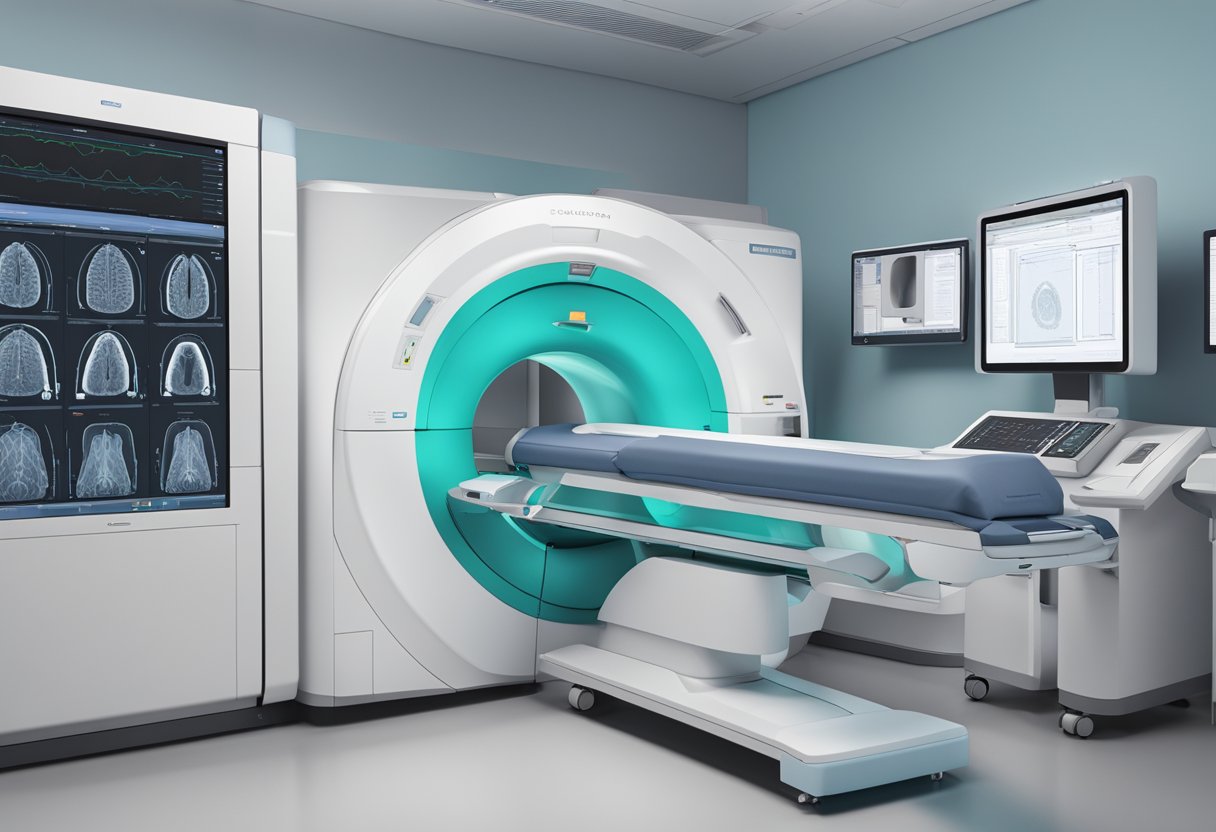
In clinical practice, identifying CTD is crucial for correct diagnosis and treatment planning. Cardiac CT and cardiovascular computed tomography (CI) are important tools for diagnosing CTD. They provide detailed images of the heart’s structure, aiding in the detection of the membrane that characterises CTD.
Guidelines established by the Society of Cardiovascular Computed Tomography (SCCT) and other related bodies are essential for the standardised use of these imaging technologies. Following these guidelines ensures accurate diagnosis, proper assessment of the related heart anomalies, and timely surgical interventions, if necessary. Additionally, standardised medical terminology, such as those provided by RadLex, assists in clear communication among healthcare professionals, improving patient outcomes.
Advancements and Protocols in Cardiac CT
In recent years, there have been significant advancements in cardiac computed tomography (CT) technology, along with the development of detailed assessment and reporting standards. Guidelines and recommendations have been established to ensure consistent and accurate results in clinical practice.
Technological Progress in Cardiac CT
Cardiac CT technology has improved significantly. Multi-energy CT and photon counting detectors allow for high-resolution images with lower radiation doses. These advancements enable detailed imaging of coronary arteries and heart structures.
Electrocardiogram (ECG) gating is a technique used to synchronise the CT scan with the heart’s ECG signal, minimising motion artifacts. Extracellular volume (ECV) measurement is another progressive feature, providing information on myocardial tissue composition.
Cardiac CT also employs iodine-based contrast agents to enhance the visibility of blood vessels, further improving the diagnostic accuracy of coronary artery disease (CAD).
Assessment and Reporting Standards
Standardised medical terminology and reporting guidelines are crucial in cardiac CT to ensure clarity and precision. The Society of Cardiovascular Computed Tomography (SCCT) and RadLex have developed consistent nomenclature for describing cardiac CT findings.
Protocols like CAD-RADS 2.0 (Coronary Artery Disease – Reporting and Data System) provide a structured reporting format, assisting clinicians in the assessment and communication of CAD severity.
Agatston score is used to quantify coronary artery calcium, indicating the extent of calcified plaque and cardiac risk. These standardised assessment tools are essential for reliable and reproducible evaluations.
Guideline and Recommendations
The development of guidelines by organisations such as the American Heart Association (AHA) and the American College of Radiology (ACR) ensures that cardiac CT practices are evidence-based and standardised.
Recommendations cover patient selection criteria, imaging protocols, and safety measures, including radiation dose management. The Society of Cardiovascular Computed Tomography (SCCT) regularly updates these guidelines to reflect new technological advancements and clinical research findings.
Adherence to these guidelines ensures high-quality care and optimised patient outcomes in cardiac CT imaging. The collaboration among these organisations reinforces the importance of a unified approach in cardiac imaging practices.
Frequently Asked Questions

Here you’ll find answers to common questions about connective tissue diseases (CTDs) in cardiology. This information can help you understand symptoms, diagnosis, life expectancy, and more.
What are typical symptoms of connective tissue diseases in women?
Women with connective tissue diseases often experience joint pain, muscle weakness, and fatigue. They may also have skin rashes or ulcers. Some may suffer from Raynaud’s phenomenon, causing cold and numb fingers or toes.
How is CTD-associated interstitial lung disease diagnosed?
Diagnosis typically involves a combination of methods. Doctors use imaging tests like high-resolution CT scans to look at lung structure. Pulmonary function tests measure how well the lungs are working, and blood tests can reveal specific antibodies related to CTDs.
What is the life expectancy for individuals with CTD-associated ILD?
Life expectancy varies and depends on the type and severity of the CTD and ILD. Early diagnosis and treatment improve outcomes. People receiving effective management for their condition often live longer and have a better quality of life.
Which diseases are included in the connective tissue diseases category?
The connective tissue diseases category includes lupus, scleroderma, rheumatoid arthritis, and polymyositis, among others. These diseases affect the proteins that support other tissues and organs in the body.
How does CTD affect the heart?
CTDs can lead to heart-related issues such as inflammation of the heart muscle or lining, increased risk of heart attacks, and problems with heart valves. Early intervention is critical to manage these complications effectively.
What are the latest guidelines for managing CTD-ILD?
Recent guidelines recommend using a multidisciplinary approach. Treatment often involves medications like corticosteroids and immunosuppressives to reduce inflammation. Regular monitoring through lung function tests and imaging helps tailor these treatments to the patient’s needs.